Scientists devise ‘glowscope’ to bring fluorescent microscopy to schools Premium
The Hindu
Students and researchers in resource-poor labs can use Foldscopes and ‘glowscopes’ to reveal more about the microscopic world.
In 2014, a group of scientists at Stanford University released Foldscope, a handheld microscope made almost entirely out of paper, which takes 30 minutes to put together, and which could capture images of cells. So far, millions of people – especially schoolchildren – around the world have taken images of the microscopic world with Foldscopes, while dozens of scientific studies have been conducted with the help of this instrument. Its cost? Rs 400.
Foldscope democratised the world’s access to optical microscopy. Now, researchers at Winona State University, Minnesota, have created a design for a ‘glowscope’, a device that could democratise access to fluorescence microscopy – at least partly so.
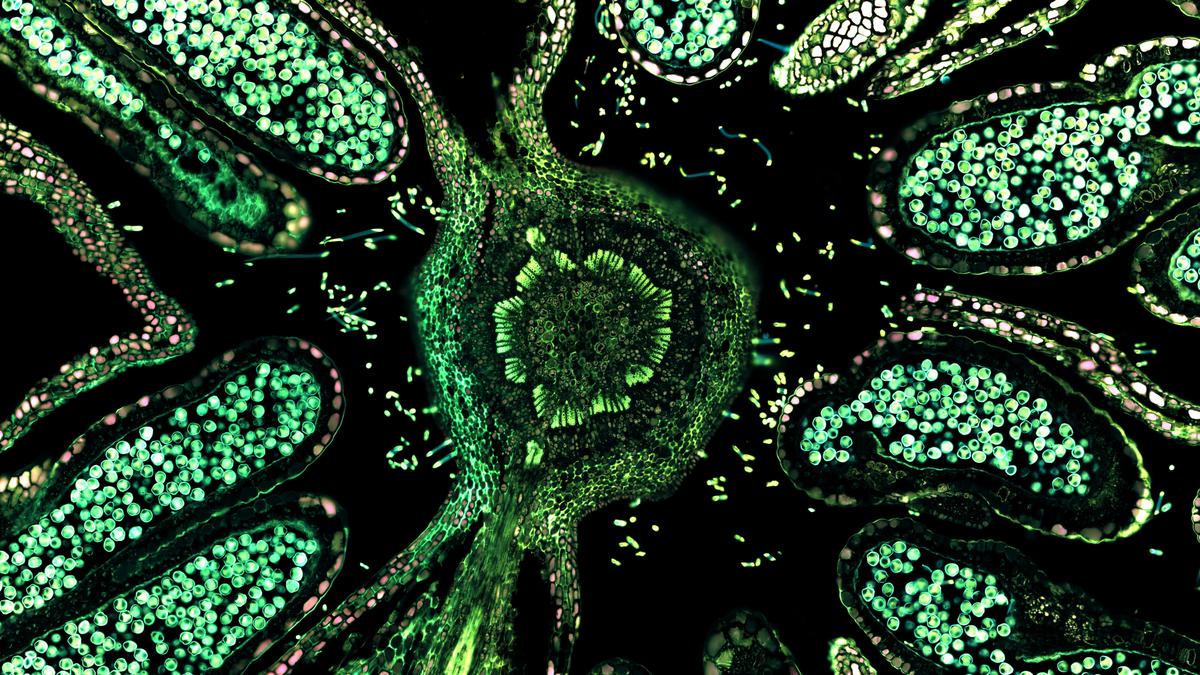
An optical microscope views an object by studying how it absorbs, reflects or scatters visible light. A fluorescence microscope views an object by studying how it reemits light that it has absorbed, i.e. how it fluoresces. This is its basic principle.
The object is illuminated with light of a specific wavelength. Particles in the object absorb this light and reemit it at a higher wavelength (i.e. different colour). These particles are called fluorophores; the object is infused with them before being placed under the microscope.
There are versions of fluorescent microscopes with more sophisticated abilities, such as epifluorescence and confocal laser-scanning microscopes.
When the fluorophores fluoresce, a fluorescent microscope can track them as they move inside the object, revealing the object’s internal shape and other characteristics. For example, a fluorophore called the Hoechst stain binds to DNA and is excited by ultraviolet light. So a tissue sample collected from a person could be injected with the Hoechst stain and placed under a fluorescent microscope. When the sample is illuminated by ultraviolet light, the stain absorbs the light and reemits it at a higher wavelength. The microscope will point out where this is happening: in the nuclei of cells, where DNA is located. This way, the nuclei in the tissue can be labelled for further study.
Scientists have developed different fluorophores to identify and study different entities, from specific parts of DNA to protein complexes. On the flip side, fluorescence microscopes cost at least a lakh rupees, but often up to crores.

Discover the Minor Planet Centre, the key hub for tracking and verifying observations of small solar system bodies.

In this week's Science for All newsletter, XX explains

Test your knowledge with the quiz on Science films at the Oscars through history

Set between the Siwalik hills and the plains, Butwal offers travellers a mix of regional cuisine, pilgrimage trails to Lumbini and the comfort of Hyatt’s new hospitality landmark

Explore 2026's wallpaper trends in India, highlighting botanicals and handmade designs that enhance modern homes with nature-inspired aesthetics.

Mercedes-Benz CLA 250+ with EQ Technology review: design, performance, range and features explained. Discover how the new electric CLA combines advanced AI, long range, fast charging and futuristic styling for the next generation of luxury EVs.

AI tool for sparrow conservation

BTS’s comeback with ‘Arirang’ marks a major global music event. When does the album release, and where can you watch their concert live stream? We have all the details

The 2026 U.S. cholesterol guidelines urge starting screening and treatment at 30 to prevent cardiovascular disease effectively.

Olam Festival 2026 in Thiruvananthapuram returns March 27-29 with 120+ vendors, music, art, and cultural zones.

A few storytellers in Thiruvananthapuram share their journeys, challenges, and evolving methods on World Storytelling Day, inspiring young minds.

76-year-old Kurian Jacob from Kerala triumphs with seven medals, including five golds, at the Open Masters Games in Abu Dhabi.

Explore the nature of the human mind as an emergent property of brain activity and complex neural interactions.